Liver Anatomy Rib Cage - Easy Notes On Liver Learn In Just 4 Minutes Earth S Lab / Learn about its function, parts, location on the body, and normally you can't feel the liver, because it's protected by the rib cage.
Liver Anatomy Rib Cage - Easy Notes On Liver Learn In Just 4 Minutes Earth S Lab / Learn about its function, parts, location on the body, and normally you can't feel the liver, because it's protected by the rib cage.. Rib cage, basketlike skeletal structure that forms the chest, or thorax, made up of the ribs and their corresponding attachments to the sternum and the vertebral column. Anatomy print anatomical organs poster brain heart lungs liver pelvis rib cage human anatomy art painting clinic wall art decor. The rib cage protects this organ. They articulate with the vertebral column posteriorly, and terminate anteriorly as cartilage (known as costal cartilage). Rib cage and internal organs.
Webmd's liver anatomy page provides detailed images, definitions, and information about the liver. However whether this gives much protection varies on the exact mechanics of injury. 'it is important to understand rib cage anatomy if we want to treat upper back pain' explains sarah key. 2 describe the anatomical and physiological/surgical lobes of liver(see the diagram above). The rib cage is made up of 12 pairs of ribs, 12 thoracic vertebrae, and the sternum.
Virtual Reality Simulation Of Liver Biopsy With A Respiratory Component Intechopen from www.intechopen.com Fundus of stomach lies posterior & lateral to left lobe of liver. Free for commercial use no attribution required high quality images. Another important feature of the rib cage is the manubriosternal joint also known as the sternal angle of louis. The rib cage protects this organ. Various skeletal muscles are attached to the rib cage. The inferior opening is the inferior thoracic aperture (anatomical thoracic outlet). The thoracic cage (rib cage) is the osteocartilaginous structure found in the axial skeleton's thoracic region. The liver (hepar) is an extremely important organ in the body of mammals and vertebrates as it provides functions essential for life.
Rib cage and internal organs.
Skeletal muscles attached to the rib cage: 'it is important to understand rib cage anatomy if we want to treat upper back pain' explains sarah key. Fashion anatomical human rib cage anatomy necklace high quality maxi vintage necklace pendants statement jewelry. The inferior opening is the inferior thoracic aperture (anatomical thoracic outlet). Find images of rib cage. Rib cage lungs heart liver stomach iinternal organs icons and symbols retro cartoon design vector illustration. Inflammation or scarring of the liver, such as with hepatitis or cirrhosis may cause rib cage pain. Structure of a typical rib: Learn also about the liver anatomy. We must be careful about what we eat. The liver has two large sections, called the right and the left lobes. They also have a role in. You know that we are all what …
It is formed by the 12 thoracic vertebrae, 12 pairs of ribs and associated costal trauma sufficient to displace these joints often injures underlying structures such as the diaphragm and/or liver, causing severe pain, particularly. The rib cage protects this organ. Another important feature of the rib cage is the manubriosternal joint also known as the sternal angle of louis. See more ideas about rib cage, human anatomy, anatomy. Rib cage, arm and eye and spine, anatomy of human bones set.
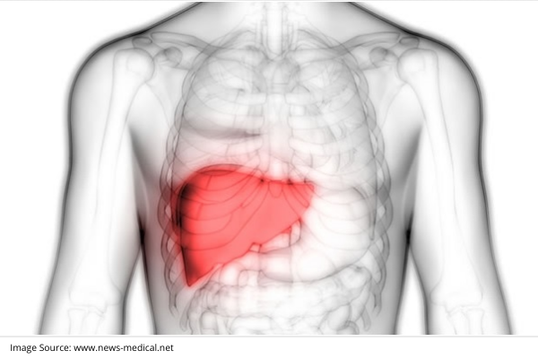
Treatment Options For Liver Cancers from www.kimshealth.org The liver has two large sections, called the right and the left lobes. 2 describe the anatomical and physiological/surgical lobes of liver(see the diagram above). Find images of rib cage. The rib cage has three important purposes : The thoracic spine supports twelve pairs of ribs that slope gently down from the back as they pass around to encase the thorax. Another important feature of the rib cage is the manubriosternal joint also known as the sternal angle of louis. We must be careful about what we eat. The liver is partially surrounded by the ribs, and extends from the level of the fifth intercostal space to the lower margin of the right rib cage, which protects this highly vascular organ from blows that could rupture it.
The liver has two large sections, called the right and the left lobes.
Rib cage anatomy and structure. Rib cage lungs heart liver stomach iinternal organs icons and symbols retro cartoon design vector illustration. It encloses and protects the heart and lungs. Another important feature of the rib cage is the manubriosternal joint also known as the sternal angle of louis. Fashion anatomical human rib cage anatomy necklace high quality maxi vintage necklace pendants statement jewelry. Rib cage, arm and eye and spine, anatomy of human bones set. Various skeletal muscles are attached to the rib cage. Human ribcage with skull sketch. Systems of man body and organs. Greater part of right lobe is covered by rib cage. The thoracic cage (rib cage) is the osteocartilaginous structure found in the axial skeleton's thoracic region. Anatomy print anatomical organs poster brain heart lungs liver pelvis rib cage human anatomy art painting clinic wall art decor. However whether this gives much protection varies on the exact mechanics of injury.
The rib cage surrounds the lungs and the heart, serving as an important means of bony protection for these vital organs. Structure of a typical rib: Learn also about the liver anatomy. 2 describe the anatomical and physiological/surgical lobes of liver(see the diagram above). Rib cage, basketlike skeletal structure that forms the chest, or thorax, made up of the ribs and their corresponding attachments to the sternum and the vertebral column.
Virtual Reality Simulation Of Liver Biopsy With A Respiratory Component Intechopen from www.intechopen.com 2 describe the anatomical and physiological/surgical lobes of liver(see the diagram above). Inflammation or scarring of the liver, such as with hepatitis or cirrhosis may cause rib cage pain. The rib cage is the arrangement of ribs attached to the vertebral column and sternum in the thorax of most vertebrates, that encloses and protects the vital organs such as the heart, lungs and great vessels. It sits above the right kidney, intestine and the stomach. Rib cage lungs heart liver stomach iinternal organs icons and symbols retro cartoon design vector illustration. They articulate with the vertebral column posteriorly, and terminate anteriorly as cartilage (known as costal cartilage). The inferior opening is the inferior thoracic aperture (anatomical thoracic outlet). Rib cage and internal organs.
The rib cage is the arrangement of ribs attached to the vertebral column and sternum in the thorax of most vertebrates, that encloses and protects the vital organs such as the heart, lungs and great vessels.
Anatomical studies have shown that the liver has one of the most complex histological structures. Fashion anatomical human rib cage anatomy necklace high quality maxi vintage necklace pendants statement jewelry. Most of left lobe is covered by rib. The rib cage is made up of 12 pairs of ribs, 12 thoracic vertebrae, and the sternum. 2 describe the anatomical and physiological/surgical lobes of liver(see the diagram above). It is of clinical importance as various structures and anatomical boundaries occur at this joint. The thoracic cage (rib cage) is the osteocartilaginous structure found in the axial skeleton's thoracic region. The heart sits atop the diaphragm and the abdominal liver is just below. Body and thorax and pelvis, extremity and femur. The gross anatomical division of the liver into its right and left lobes is useful in gross description but is without morphological signicance. Greater part of right lobe is covered by rib cage. However whether this gives much protection varies on the exact mechanics of injury. Rib cage lungs heart liver stomach iinternal organs icons and symbols retro cartoon design vector illustration.
The thoracic cage (rib cage) is the osteocartilaginous structure found in the axial skeleton's thoracic region anatomy rib cage. The areas of supply of the right and left hepatic arteries, with accompanying portal vein and bile duct branches, can be demarcated by a line passing through the.


Posting Komentar
0 Komentar